For pet owners searching for “animal xray and ultrasound near me,” understanding how imaging helps in surgery is very important. X-rays and ultrasounds give vets a clear look at bones, organs, and tissues. This helps them plan surgery more safely and carefully. Accurate imaging lets vets find the exact problem, reduce risks, and make sure your pet heals comfortably. Using these tools also lowers stress for pets and owners, since the vet can explain the procedure with clear visuals.
How Imaging Helps Surgeons Make Decisions
X-rays show bones and joints in detail, while ultrasounds let vets see soft tissues and organs in real time. Using both tools gives a full picture of the problem before surgery. For example, vets can see broken bones, bladder stones, or lumps in the abdomen. Knowing the details helps them decide the best surgery method, reduce mistakes, and improve recovery.
In more complex cases, imaging allows vets to measure the size of a tumor or the depth of a bone fracture. This precision ensures that surgical tools are chosen correctly and that the surgery plan is specific to the pet’s needs.
Finding Hidden Problems Early
Some health issues cannot be felt or seen from the outside. Ultrasounds can show fluid, small lumps, or organ problems that are hard to detect. X-rays can reveal hidden fractures or unusual bone shapes. Finding these problems before surgery allows to treat everything at once. This keeps pets from needing more surgeries later.
For example, a dog may be brought in for a routine operation, but imaging could reveal a small kidney cyst that needs attention. Addressing it during the same procedure prevents extra stress and additional anesthesia for the pet.
Planning Surgery for Each Pet
Every pet’s problem is different, and imaging helps vets make a plan that fits. A cat with a soft tissue lump may need a different approach than a dog with a broken bone. Ultrasounds show the exact location of organs or lumps, and X-rays show the shape and alignment of bones. Planning surgery this way reduces operation time, causes less stress to the pet, and helps healing happen faster.
Imaging also helps in choosing whether minimally invasive surgery or traditional surgery is better. Minimally invasive techniques often mean smaller cuts, less pain, and quicker recovery. By seeing the problem clearly, vets can choose the safest and most effective method.
Keeping Surgery Safe with Anesthesia
Surgery usually requires anesthesia, and knowing how healthy the organs are is very important. Ultrasounds can check the heart, liver, and kidneys. X-rays can show lungs and chest health. This information helps vets give the right amount of anesthesia safely, so pets stay safe and comfortable during surgery.
Pets with heart or lung conditions can be monitored more carefully because the vet already knows the state of these organs from imaging. This reduces the risk of complications during surgery and ensures a smoother procedure.
Checking Recovery After Surgery
Imaging continues to help even after surgery. Follow-up X-rays and ultrasounds show if bones, tissues, and organs are healing properly. They also help detect infections or other problems early. This allows vets to adjust care and help pets recover faster and fully.
For instance, after orthopedic surgery, X-rays can confirm that a bone is healing correctly. Ultrasounds can check internal organs for any signs of fluid buildup or inflammation. This gives owners peace of mind that their pet is on track to full recovery.
Helping Pet Owners Understand
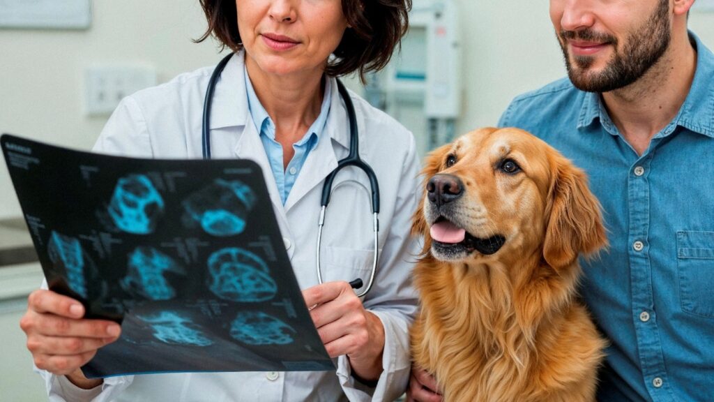
X-rays and ultrasounds show pictures of the pet’s problem, making it easier for owners to understand. Seeing the images helps owners follow care instructions correctly and feel confident about the treatment. This also helps owners notice any changes in their pet’s health quickly.
Some owners find it reassuring to see a clear image of their pet’s problem. It makes the surgery seem less intimidating and helps them understand why certain steps in post-operative care, such as rest or medication, are important.
Final Look:
At Fraser MacDonald Animal Hospital, we use X-rays and ultrasounds to plan surgery carefully and give pets the safest care. With clear imaging and skilled veterinary care, our team can target the problem accurately and reduce risks. We also guide owners on care after surgery to help pets heal well, including tips for dog umbilical hernia surgery recovery.
Take action today: schedule a visit with Fraser MacDonald Animal Hospital to give your pet safe and accurate surgical care using advanced X-ray and ultrasound technology. Our team is ready to provide personalized attention and expert guidance to keep your pet healthy and comfortable.
FAQs:
X-rays show bone structure and alignment, helping vets plan surgery accurately and safely.
Ultrasounds reveal soft tissues, organs, and fluid issues, allowing vets to identify hidden problems.
Yes, it helps veterinarians plan precisely, minimizing complications and improving recovery outcomes.
They are used post-surgery to monitor healing, check for infections, and ensure proper recovery.
Yes, imaging assesses organ health, helping vets adjust anesthesia for safe and comfortable surgery.